
Cells move material by wrapping it in membrane bubbles, or vesicles, that travel to new locations. These vesicles merge with other vesicles to deliver their contents, a process that requires 2 membranes to connect without tearing or leaking. Scientists have long proposed that merging cell membranes undergo a short-lived halfway state during this process, but none had previously been able to directly visualize it inside intact cells.
Researchers from NIH and the University of Virginia set out to test whether membranes in living cells form a stable, observable structure that represents this halfway state when they merge. They grew several types of mammalian cells, including human, monkey, mouse, and rat cells, in laboratory culture flasks filled with a nutrient-rich solution. They kept the flasks inside an incubator set to 37°C (98.6°F) to keep the cells alive and growing.
The researchers placed between 80,000 and 100,000 cells onto tiny gold-coated platforms designed for high-resolution imaging. To preserve cellular structures in their native state, the team rapidly froze the cells to lock their membranes in place. Then they examined the frozen cells from multiple angles using a method called cryo-electron tomography, which produces images called tomograms.
The team used these tomograms to reconstruct 3D views of the cells at the nanometer scale, which is small enough to see the thin membranes of internal vesicles and the outer cell membrane, called the plasma membrane. They created approximately 300 3D reconstructions of regions near the edge of the cells, where membrane bubbles move and interact. Then they focused on membrane contact sites where 2 vesicles or a vesicle and the cell’s plasma membrane came into proximity.
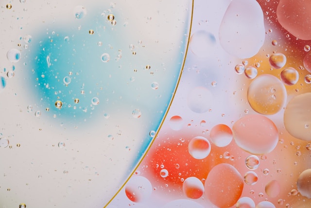
Typical cell membranes are built from 2 separate layers of fat-like molecules that form a flexible barrier. However, these researchers identified a previously uncharacterized membrane structure that formed when the outer layers of 2 membranes merged into a continuous sheet while the inner layers remained separate. Where the 2 membrane vesicles touched, the researchers saw a flat, circular path where the outer layers of the membranes joined together. This patch formed at the contact point between the 2 vesicles, creating a thin membrane bridge between them, similar to the film that forms where 2 soap bubbles press against each other. The scientists called this configuration a hemifusome.
The researchers explained that the hemifusomes were far larger and more stable than the short-lived halfway states proposed by earlier researchers. They interpreted this long-term stability to mean that hemifusomes may persist long enough to perform cellular functions rather than representing only transient fusion events.
They also saw that some hemifusomes contained a single lens-shaped droplet sitting within the membrane at the contact point where the 2 vesicles had partially fused. The researchers observed these droplets in approximately half of the 308 tomograms they examined. The droplets were on average 40 nanometers across, about 100 times smaller than the surrounding vesicles, and in contact with the oily interior of the membrane.
The droplets looked different from the surrounding membrane lipids, suggesting that they contain a mixture of lipids and proteins. The scientists referred to this mixed structure as a proteolipid nanodroplet. They interpreted the consistent one-to-one association between a hemifusome and its single proteolipid nanodroplet to suggest that the nanodroplets may help stabilize the hemifusome or contribute to changes in the shape and organization of cell membranes.
To test whether the hemifusomes helped cells move materials around, the researchers placed 5- or 15-nanometer-sized particles of gold into the cells. These particles were small enough to move through the cell’s internal transport system, which normally carries nutrients and other molecules. Using powerful microscopes, the team followed the gold particles as they passed through the cell’s transport compartments. However, the particles never entered the hemifusomes, indicating that they’re probably not involved in cellular transport.
The researchers concluded that hemifusomes form when cell membranes join together or reshape themselves, like temporary construction sites where cell membranes are built, repaired, or rearranged. Unlike current models of membrane fusion and vesicle formation, these results suggest that key intermediate states could form stable, functional cellular structures.
The researchers proposed that future workers should identify the molecular composition of the proteolipid nanodroplets and determine how cells regulate the transition from hemifusomes to fully fused membranes. They also suggested investigating whether hemifusomes participate in vesicle formation, membrane recycling, or stress responses in these or other cell types.
