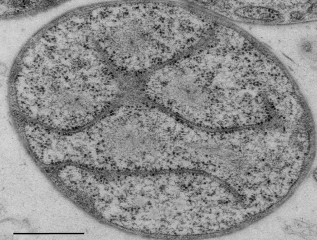
When we first learn about cells in biology, we are taught that bacterial cells are simple, and animal and plant cells are more complex. Mammalian cells, the cells that make up dogs, humans and all mammals, are “eukaryotes.” Eukaryotes have multiple membranes — one surrounding their innermost section, the nucleus, which protects their DNA and another outer membrane enclosing the entire cell. Bacterial cells, also called “prokaryotes,” have only an outer membrane.
However, reality is never so black and white! As it turns out, there are some species of bacteria that have a more complex membrane system resembling the animal-like cells (eukaryotes). One example of these more complex bacteria is Gemmata obscuriglobans. These bacteria are aquatic and can be found in fresh or brackish bodies of water, such as lakes and ponds. Although there has been some debate on this subject, a recent paper in PLOS One has cleared up the controversy about this membrane system with a rigorous study.
Using three different techniques, scientists at the University of Queensland broke apart, or lysed, some G. obscuriglobans cells — freezing the cells with liquid nitrogen (at a frigid -321 deg F) and grinding them up, making thin slices of the cells. These small slices are then stained and highlights the details of the structure of the cell. After lysing the cells, the different slices were viewed under a powerful electron microscope. An electron microscope captures far more detail at much higher magnitude than a traditional light microscope you may see in schools.
After analysing the cell slices to examine the structure, the researchers found three distinct membrane types. They confirmed that these different types of membrane enclose separate sections of the cell. Before this study, it was previously thought of this particular bacteria that the membranes may appear separate, but were really just a continuous membrane with an unusual shape, make it appear as three separate ones. This created a mystery, because certain processes, such as the process that reads and duplicates DNA, only occur outside the innermost membrane. Therefore, the process only occurring in certain parts of the cell indicated that there were true separate compartments. Also, one of the three membrane types contained structures called nuclear pores. Nuclear pores are channels in the membrane that allow large molecules, like nutrients or proteins, to pass through. This result is exciting because many eukaryotic cells have nuclear pore structures, but bacteria usually don’t.
Researchers were even able to determine the size and structural characteristics of the nuclear pore. The size and layout of the nuclear pores closely resemble yeast cells, plant cells, and human liver cells. One might wonder if that means that these unique bacteria might be distantly related to the eukaryotic cell line. Relationships like this can be traced by comparing the genetic code of each organism to see how similar they are. However, it turns out that instead of evolving from a eukaryotic cell, this might actually be an example of convergent evolution. Convergent evolution is the event where two completely separate ancestries of different living things end up with the same characteristics despite starting off differently.
This is a profound conclusion from an evolutionary perspective. For multiple cell membranes to occur in two separate lines of evolution (bacteria and mammals) means that this type of membrane system is a very effective way for the cell to accomplish nutrient transport.


