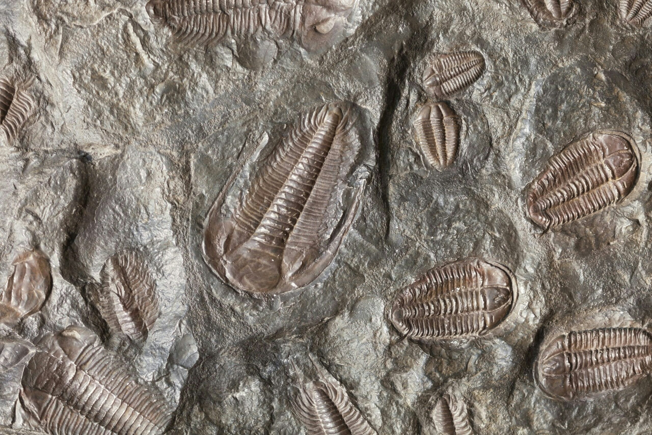

Trilobites were a diverse group of marine animals that lived from 540 to 250 million years ago. They were some of the earliest known and longest-lived arthropods. Trilobites are named after their body shape, which featured a hard exoskeleton divided into 3 lobes.
Paleontologists have described more than 20,000 different species of trilobites to date, with many different lifestyles and feeding behaviors. Some burrowed into the seafloor, while others floated freely or swam in the ocean. But everything scientists know (or think they know) about what trilobites ate comes from indirect evidence, like their gut shapes and sizes. Researchers have never found a fossilized trilobite with a full stomach. Until now…
A group of researchers from the Czech Republic and Sweden recently described a complete fossil of the trilobite Bohemolichas incola with its gut contents intact. They found this unique specimen in the Šárka Formation, in the Prague Basin of the Czech Republic. It died belly-up on the seafloor 465 million years ago and was rapidly encased in a lump of silica, known as a nodule. The researchers explained the silica nodule kept the carcass from being squished as it was buried, preserving the whole fossil in 3 dimensions for millions of years.
The team used a 3D imaging method called microtomography to peer inside the trilobite’s gut. They used this method to create a stacked set of images of the inside of the fossil, slice by slice, which computer programs knitted together into a 3-dimensional shape. Scientists traditionally use X-rays in microtomography, but this team used a specialized energy source, synchrotron radiation, to increase the resolution and contrast of the images. Synchrotron radiation is high-intensity light produced by electrons moving at nearly the speed of light in a circular accelerator called a synchrotron. They combined this method with another type of imaging, known as propagation phase contrast imaging, to further enhance the contrast between soft tissues that absorb regular light similarly.
From these images, the researchers found the trilobite’s gut was entirely full of crushed bits of shell made of calcium carbonate. They identified the shells as mostly from small crustaceans about the size of ants, called ostracods. Some of the shell bits came from larger 2-shelled creatures, similar to clams or mussels, and others were from a single organism resembling a starfish. All these critters lived in mud at the bottom of the ocean, suggesting the trilobite fed on them as it scurried along the seafloor. Since the trilobite ate several different types of shelly creatures, the researchers inferred it was a scavenger that indiscriminately hoovered up whatever it came across, rather than a predator that selectively hunted its prey.
The team also noted the shells within the trilobite’s gut had sharp edges and no signs of etching. They interpreted this to mean the trilobite’s digestive tract had a neutral or alkaline pH, because the shells would have begun to dissolve if the gut was acidic, like in humans and most mammals. The researchers explained the enzymes that help animals digest food are very sensitive to pH. Hence, this evidence suggests the trilobite had enzymes similar to those of other creatures with neutral or alkaline digestive systems. Living examples of these organisms include crustaceans, like shrimp and lobsters, and chelicerates, like spiders and scorpions.
Finally, the researchers discovered a set of tiny tunnels burrowing into the trilobite’s remains, indicating the creature fell victim to its own set of scavengers, after it died but before it was encased in silica. They found the most concentrated set of burrows near the trilobite’s head, which appeared to be the most intense area of feeding. They also found some burrows in the lower part of the trilobite’s body, but none of them went into its digestive tract, meaning the scavengers avoided the animal’s gut entirely. The researchers suggested the gut could have remained toxic for some time if its enzymes continued to digest the animal’s final meal after it died.
The researchers concluded the 3D specimen of Bohemolichas incola they described provides the best knowledge to date about the feeding habits of trilobites, including what they ate and how they digested it. They also suggested this particular trilobite’s physiology could mean a near-neutral-pH gut was characteristic of the most primitive arthropods. However, they also pointed out very few scientists have studied how intestinal pH affects digestion in living arthropods, so more work is needed to test this hypothesis.
