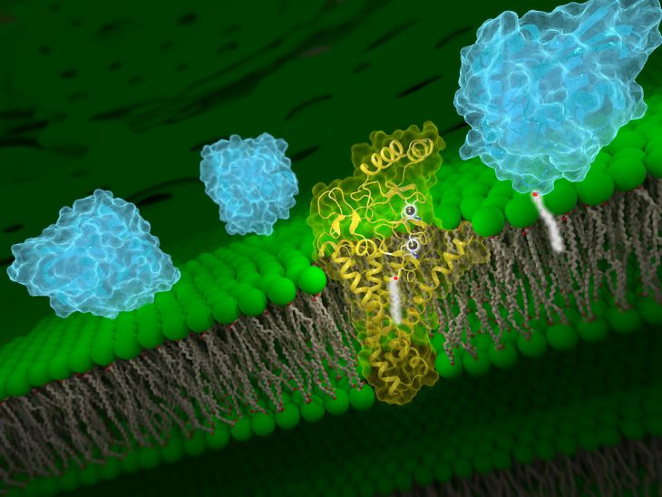
Infectious pathogens such as viruses and bacteria are a constant threat to our health and well-being. However, our bodies resist these attackers through a complex immune system made of many different cell types producing many different proteins. For this network of cells to work properly, they must circulate throughout the body and interact with other members of the immune system. Most of these interactions are mediated through a collection of hundreds of different proteins that are on the outside of the cell membrane.
Scientists know that membrane proteins help the different immune cells work together to control infections, identify tumors, and even cause autoimmune diseases. But they don’t quite understand how they do this. A team of researchers from the UK and Switzerland recently designed a large-scale experiment to use the presence and absence of different membrane proteins on immune cells across the body to create a network showing these many interactions.
The team identified 630 different membrane proteins that are produced by immune cells. To study each membrane protein, the team used a piece of DNA to force the cells to make a lot of a single membrane protein at a time. The team then collected the produced proteins and coated them with another protein called biotin. A portion of these biotin-coated proteins were attached to the bottom of a special plastic dish.
One-by-one, each attached protein was mixed with a biotin-coated protein for a few minutes. If the two membrane proteins are able to interact with each other, then they bind to one another. By flooding these plates out with a watery solution, any proteins that did not bind to the plate-attached protein would be washed away. Lastly, a glowing molecule was added to the plates that can only bind to a biotin-coated protein. If a pair of proteins could interact, the glowing molecule would stay in the plate and could be seen by a powerful microscope. With this, the team could detect whether two membrane proteins interact or not.
By comparing every possible combination of the 630 membrane proteins this way, the team verified nearly all of the previously known pairs and identified 28 new pairs. The strength of these interactions was measured by a process called surface plasmon resonance. In a new tube, the team mixed together two proteins that could interact and fired a beam of electrons at the mixture. When proteins would bind, they would alter the beam a little bit. By adding proteins together slowly, the team could measure how much protein is needed for binding to happen and for the beam to change. This measurement also told the team how strong the bond between two proteins is.
With these measurements, the team made a network of each protein interaction and how strong that interaction was. With this information, the network showed which proteins interact with which. Additionally, when they found that one protein could interact with several others, the researchers could predict which it would prefer based on the interaction strength.
While the network the team made shows the strength of membrane protein interactions, not every cell will produce every membrane protein. The team turned to past work on membrane proteins to find the amounts and types of proteins that are expressed and how these change with different locations in the body. With this final piece of information, the network now shows which cells interact with which, how tightly these interactions happen, and can ultimately predict how immune cells from one part of the body will work with cells from another part.
With this network, the team hopes that medical professionals and scientists can make more informed decisions about healthcare and further research into immune health such as the progression of autoimmune disease. Additionally, the team points out that the 28 new interactions have not previously been studied in living cells and need to be verified. Studying where these proteins are expressed in the body and what cells are using them may lead to a more complete picture of the immune system.


